
Health Library
Palpebral slant - eye
Mongolian slant
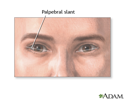
The palpebral slant is the direction of the slant of a line that goes from the outer corner of the eye to the inner corner.
Images
I Would Like to Learn About:
Considerations
The word palpebral refers to the upper and lower eyelids, which help determine the shape of the eye. A line drawn from the inner corner to the outer corner determines the slant of the eye, or palpebral slant. Slanting and a fold of skin (epicanthal fold) are normal in people of Asian descent.
Abnormal slanting of the eye may occur with some genetic disorders and syndromes. The most common of these is Down syndrome. People with Down syndrome often also have an epicanthal fold in the inner corner of the eye.
Causes
Palpebral slant may not be due to any defect. However, in some cases, it may be due to:
- Down syndrome
- Fetal alcohol syndrome
- Certain genetic disorders
When to Contact a Medical Professional
Contact your health care provider if:
- Your infant has abnormal features of the face
- You are worried about your infant's ability to move their eyes
- You notice any abnormal color, swelling, or discharge from your infant's eyes
What to Expect at Your Office Visit
Your provider will perform a physical exam and ask questions about your child's medical history and symptoms.
An infant with an abnormal palpebral slant that is due to a problem will usually have other symptoms of another health condition. That condition will be diagnosed based on a family history, medical history, and a physical exam.
Tests to confirm a disorder may include:
- Chromosome studies
- Enzyme assays
- Metabolic studies
- X-rays
Related Information
Epicanthal foldsReferences
Kataguiri P, Kenyon KR. Corneal and external eye manifestations of systemic disease. In: Yanoff M, Duker JS, eds. Ophthalmology. 6th ed. Philadelphia, PA: Elsevier; 2023:chap 4.25.
Madan-Khetarpal S, Arnold G, Ortiz D. Genetic disorders and dysmorphic conditions. In: Zitelli BJ, McIntire SC, Nowalk AJ, Garrison J, eds. Zitelli and Davis' Atlas of Pediatric Physical Diagnosis. 8th ed. Philadelphia, PA: Elsevier; 2023:chap 1.
Orge FH. Examination and common problems in the neonatal eye. In: Martin RJ, Fanaroff AA, Walsh MC, eds. Fanaroff and Martin's Neonatal-Perinatal Medicine. 11th ed. Philadelphia, PA: Elsevier; 2020:chap 95.
Slavotinek AM. Dysmorphology. In: Kliegman RM, St. Geme JW, Blum NJ, Shah SS, Tasker RC, Wilson KM, eds. Nelson Textbook of Pediatrics. 21st ed. Philadelphia, PA: Elsevier; 2020:chap 128.
BACK TO TOPReview Date: 4/25/2023
Reviewed By: Charles I. Schwartz, MD, FAAP, Clinical Assistant Professor of Pediatrics, Perelman School of Medicine at the University of Pennsylvania, General Pediatrician at PennCare for Kids, Phoenixville, PA. Also reviewed by David C. Dugdale, MD, Medical Director, Brenda Conaway, Editorial Director, and the A.D.A.M. Editorial team.
 | A.D.A.M., Inc. is accredited by URAC, for Health Content Provider (www.urac.org). URAC's accreditation program is an independent audit to verify that A.D.A.M. follows rigorous standards of quality and accountability. A.D.A.M. is among the first to achieve this important distinction for online health information and services. Learn more about A.D.A.M.'s editorial policy, editorial process and privacy policy. A.D.A.M. is also a founding member of Hi-Ethics. This site complies with the HONcode standard for trustworthy health information: verify here. |
The information provided herein should not be used during any medical emergency or for the diagnosis or treatment of any medical condition. A licensed medical professional should be consulted for diagnosis and treatment of any and all medical conditions. Links to other sites are provided for information only -- they do not constitute endorsements of those other sites. No warranty of any kind, either expressed or implied, is made as to the accuracy, reliability, timeliness, or correctness of any translations made by a third-party service of the information provided herein into any other language. © 1997- 2024 A.D.A.M., a business unit of Ebix, Inc. Any duplication or distribution of the information contained herein is strictly prohibited.
![]()